neuraddiagnostic.com

Author name: Neurad Diagnostic

CT Guided Biopsy in Delhi — Accurate Tissue Diagnosis by Interventional Radiology at Neurad Diagnostics
Blog

CT Guided Biopsy in Delhi — Accurate Tissue Diagnosis & FNAC | Neurad Diagnostics

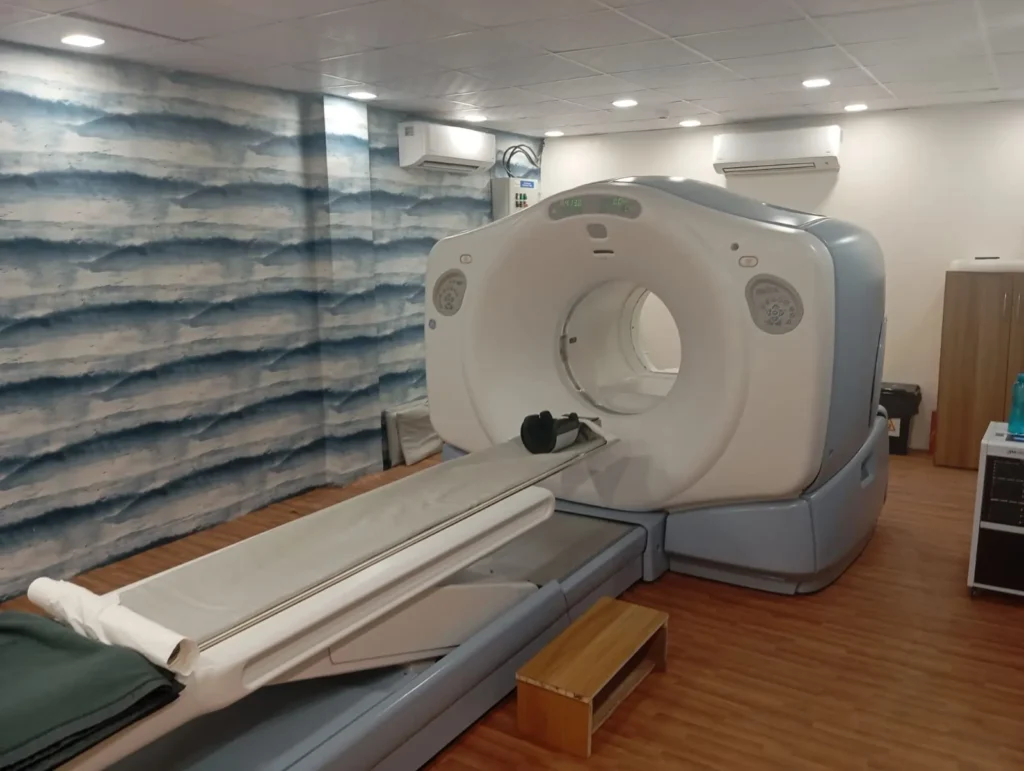
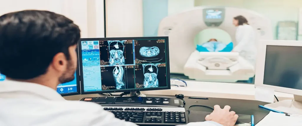
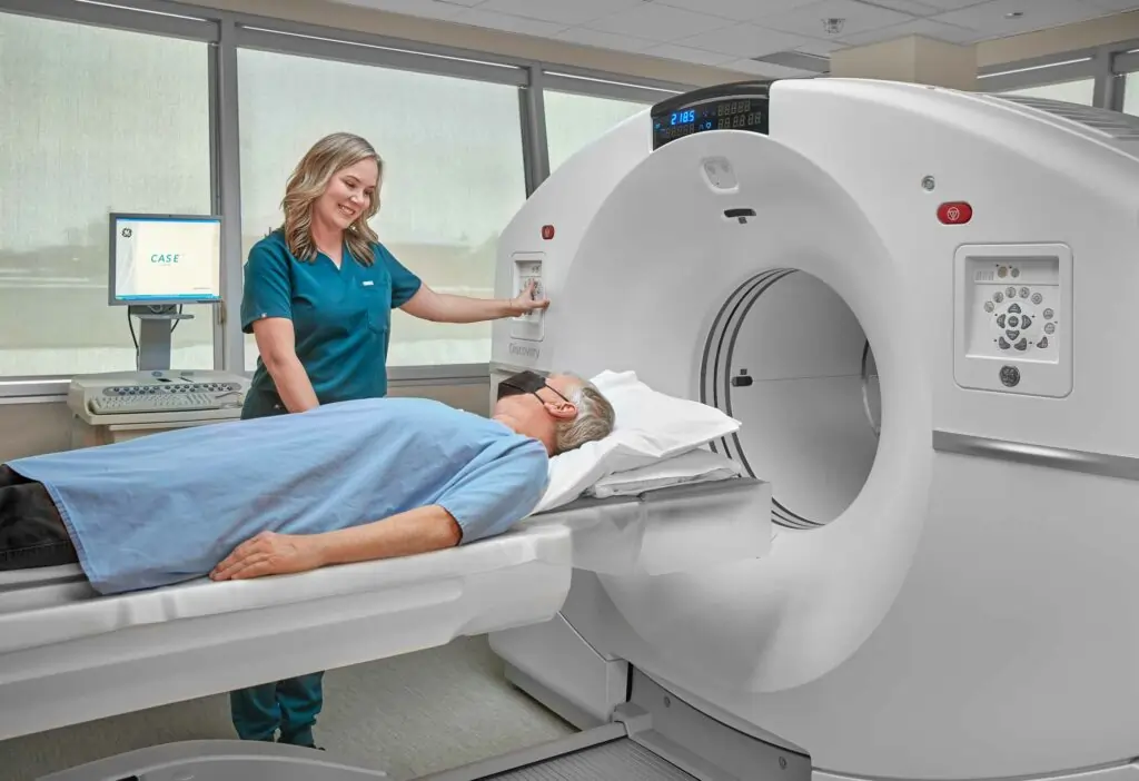
When imaging studies reveal a suspicious lesion or mass in the body, the definitive next step is usually a biopsy — obtaining a tissue sample for pathological analysis. The CT Guided Biopsy, available at Neurad Diagnostics in Delhi, is one of the most accurate, safe, and minimally invasive methods for obtaining tissue from lesions deep inside the body that cannot be reached safely by other means. Whether it is a lung nodule, liver mass, abdominal lymph node, or spinal lesion, this procedure allows our expert interventional team to deliver a precise needle to the exact target — without major surgery. This guide explains everything patients need to know about the biopsy and FNAC in Delhi. According to the Society of Interventional Radiology (SIR), CT guided biopsy is a safe and highly effective procedure for tissue diagnosis across all body regions. The Radiological Society of North America (RSNA) endorses image-guided biopsy as the standard of care for accessible lesion sampling. this test at Neurad Diagnostics Delhi — Precision tissue sampling under CT guidance Table of Contents What Is core needle biopsy? this procedure vs. CT Guided FNAC Why Doctors Recommend CT Guided Biopsy Conditions Diagnosed by this test Who Needs CT Guided Biopsy? Symptoms and Findings That Lead to the procedure How CT Guided Biopsy Works Preparation Before this procedure Step-by-Step the biopsy Procedure at Neurad Diagnostics Advantages of CT Guided Biopsy Accuracy and Success Rate Safety and Complication Profile Cost of CT Guided Biopsy in Delhi NCR Why Choose Neurad Diagnostics for CT Guided Biopsy in Delhi? Frequently Asked Questions About this test 1. Is CT guided biopsy the same as a surgical biopsy? 2. Can CT guided biopsy be done for any body part? 3. Will I need to stop blood thinners before the biopsy? 4. How long will I be at the hospital/center for this procedure? 5. When will I get the biopsy results? 6. Is the biopsy suitable for lung nodules? 7. Can this test diagnose tuberculosis? 8. Does CT guided biopsy cause cancer to spread? 9. Can I eat after the procedure? 10. Where can I get this procedure near me in Delhi NCR? Conclusion — Book Your the biopsy at Neurad Diagnostics Delhi What Is core needle biopsy? CT Guided Biopsy is an interventional radiology procedure in which real-time CT (Computed Tomography) imaging is used to precisely navigate a biopsy needle to a target lesion within the body. The interventional radiologist uses serial CT images to visualize the exact position of the needle tip in relation to the target lesion and surrounding critical structures, adjusting its trajectory in real time until the needle is optimally positioned. A tissue core sample (biopsy) or fine needle aspirate (FNAC) is then obtained from the lesion for pathological analysis. CT guidance is particularly valuable for accessing lesions that are deep within the body, are not visible on ultrasound, are located near critical structures (blood vessels, nerves), or require very precise needle placement for safety. Lesions in the lung, liver, pancreas, kidney, adrenal gland, retroperitoneum, spine, mediastinum, and bone are commonly biopsied under CT guidance. At Neurad Diagnostics in Delhi, our experienced interventional team performs the procedure as a same-day, outpatient or short-stay procedure. this procedure vs. CT Guided FNAC Two types of needle sampling are performed under CT guidance at Neurad Diagnostics: CT Guided FNAC (Fine Needle Aspiration Cytology) uses a very thin needle (22-25 gauge) to aspirate a small number of cells from a lesion. It is less invasive and carries a lower bleeding risk. However, it provides only cytological (cellular) material, which may be insufficient for certain diagnoses requiring tissue architecture assessment. CT Guided Core Biopsy (Trucut Biopsy) uses a larger cutting needle (14-20 gauge) to obtain a core of tissue that preserves histological architecture. This provides a richer tissue sample suitable for histopathology, immunohistochemistry (IHC), molecular profiling, and genomic testing — all increasingly essential for modern targeted cancer therapy selection. The choice between FNAC and core biopsy depends on the lesion location, size, patient factors, and clinical requirements. Why Doctors Recommend CT Guided Biopsy Oncologists, pulmonologists, hepatologists, and other specialists refer patients for the biopsy in Delhi because it is the most reliable way to establish a definitive tissue diagnosis without major surgery. In the modern era of cancer treatment, accurate tissue diagnosis is essential not just to confirm malignancy, but to identify: Cancer type and subtype (adenocarcinoma vs. squamous cell carcinoma vs. SCLC, etc.) Immunohistochemistry (IHC) markers (ER/PR/HER2 for breast cancer, PDL-1 for immunotherapy) Molecular mutations (EGFR, ALK, ROS1, KRAS, BRAF, etc.) for targeted therapy NGS (Next Generation Sequencing) profiling for comprehensive genomic analysis Lymphoma subtyping requiring tissue architecture Non-malignant diagnoses including infections (TB, fungal), sarcoidosis, and autoimmune conditions Conditions Diagnosed by this test Lung Cancer: Lung nodules, masses, and mediastinal lymph nodes Liver Lesions: Hepatocellular carcinoma, liver metastases, cholangiocarcinoma, benign lesions Pancreatic Cancer: Pancreatic masses and lymph nodes Kidney Tumors: Renal cell carcinoma vs. benign oncocytoma Adrenal Lesions: Adrenal metastases vs. adenoma Lymphoma: Lymph node and extranodal lymphoma Bone Lesions: Primary bone tumors and bone metastases Retroperitoneal Masses Spinal Lesions Soft Tissue Masses Infections: Abscess characterization and culture, tuberculosis Carcinoma of Unknown Primary Who Needs CT Guided Biopsy? Your physician may refer you for core needle biopsy in Delhi if: CT, MRI, or PET scan has revealed a suspicious lesion that requires tissue confirmation A lesion is too deep to be biopsied safely by physical examination or ultrasound You have a known cancer and a new lesion has appeared that needs tissue characterization Cancer has returned and repeat biopsy is needed for molecular profiling You need a diagnosis before starting chemotherapy, targeted therapy, or immunotherapy A pulmonary nodule requires tissue diagnosis to determine if it is malignant You have a liver lesion where the diagnosis is uncertain on imaging alone Suspected lymphoma requiring lymph node biopsy Symptoms and Findings That Lead to the procedure Unexplained weight loss with suspicious mass found on imaging Persistent cough or hemoptysis with lung mass on CT Abdominal

Trivehexin PET Scan in Delhi — Integrin αvβ6 Cancer Imaging at Neurad Diagnostics NCR
Blog

Trivehexin PET Scan in Delhi — Integrin αvβ6 Cancer Imaging | Neurad Diagnostics

The landscape of cancer imaging is continuously evolving, and one of the most exciting recent advances is the Trivehexin PET Scan. Available at Neurad Diagnostics in Delhi, the 68Ga-Trivehexin PET/CT scan represents a breakthrough in integrin-targeted molecular imaging for cancer. By targeting integrin αvβ6 — a protein highly overexpressed in many epithelial cancers but absent in normal adult tissues — the this scan provides a uniquely specific and high-contrast method for cancer detection, staging, and treatment planning. This guide explains everything you need to know about this innovative diagnostic technology. Research published in leading journals endorsed by the Society of Nuclear Medicine and Molecular Imaging (SNMMI) demonstrates the high potential of integrin-targeted PET imaging. The European Association of Nuclear Medicine (EANM) actively supports research in novel molecular imaging tracers including Trivehexin for cancer detection. this imaging test at Neurad Diagnostics Delhi — Advanced integrin-targeted cancer imaging Table of Contents What Is a Trivehexin PET Scan? Why Doctors Recommend the Trivehexin PET Scan Cancers and Conditions Detected by Trivehexin PET Scan Who Needs a Trivehexin PET Scan? Symptoms That May Lead to Trivehexin PET Scan How the this imaging test Procedure Works Preparation Before the the Trivehexin scan Step-by-Step Trivehexin PET Scan Procedure at Neurad Diagnostics Advantages of Trivehexin PET Technology Accuracy and Clinical Benefits Safety Information Cost of Trivehexin PET Scan in Delhi NCR Why Choose Neurad Diagnostics for Trivehexin PET Scan in Delhi? Frequently Asked Questions About this scan 1. What is integrin αvβ6? 2. Is Trivehexin PET better than FDG PET for pancreatic cancer? 3. Can Trivehexin PET detect peritoneal metastases? 4. Which lung cancer type benefits from Trivehexin PET? 5. What is the theranostic use of Trivehexin? 6. Is Trivehexin PET available in India? 7. How does Trivehexin compare to FAPI PET? 8. Is Trivehexin PET safe with chemotherapy? What Is a Trivehexin PET Scan? The the Trivehexin scan, formally known as 68Ga-Trivehexin PET/CT, is an advanced nuclear medicine imaging procedure using a Gallium-68 labelled peptide tracer called Trivehexin — a highly specific ligand engineered to bind to Integrin αvβ6 (alpha-v beta-6 integrin). Integrin αvβ6 is a cell surface receptor that plays a critical role in cancer cell invasion, proliferation, and immune evasion. Crucially, αvβ6 is normally absent or expressed at very low levels in healthy adult epithelial tissues but is massively upregulated in a wide range of epithelial cancers — including pancreatic, lung, colorectal, gastric, cervical, ovarian, and head and neck cancers. This highly cancer-specific expression pattern of αvβ6 makes Trivehexin an exceptionally promising diagnostic target. Because normal adult organs show minimal αvβ6 expression, background signal on the Trivehexin PET image is very low, allowing tumor sites to stand out with excellent contrast. Neurad Diagnostics is among the pioneering centers in Delhi NCR bringing this next-generation integrin imaging technology to patients across India. Why Doctors Recommend the Trivehexin PET Scan Oncologists recommend this PET/CT primarily in clinical situations where conventional imaging provides incomplete or misleading staging information. A key scenario is pancreatic cancer — one of the most challenging cancers to image accurately. Pancreatic ductal adenocarcinoma expresses extremely high levels of αvβ6, and Trivehexin PET has been shown to outperform FDG PET for pancreatic cancer staging, particularly for identifying peritoneal metastases and small lymph node involvement that change resectability assessment. Similarly, for colorectal cancer peritoneal metastases — a critical staging question that determines whether patients are eligible for cytoreductive surgery — Trivehexin PET’s very low abdominal background makes it particularly powerful. For lung adenocarcinoma, which uniformly expresses αvβ6, Trivehexin PET provides precise staging and can distinguish malignant from benign nodules with high confidence. Additionally, αvβ6 expression on cancer cells is being actively investigated as a target for αvβ6-directed radionuclide therapy, making Trivehexin PET a potential theranostic partner for emerging αvβ6-targeted treatments. Cancers and Conditions Detected by Trivehexin PET Scan The the procedure is applicable across multiple αvβ6-expressing cancer types: Pancreatic Ductal Adenocarcinoma: Very high αvβ6 expression — often superior to FDG PET for staging Lung Adenocarcinoma: Uniform αvβ6 expression makes Trivehexin highly effective Colorectal Cancer: Particularly useful for peritoneal metastasis detection Gastric Cancer: High αvβ6 expression in gastric adenocarcinoma Cervical Cancer: αvβ6 strongly expressed in cervical squamous and adenocarcinoma Ovarian Cancer: Useful for peritoneal spread assessment Head and Neck Squamous Cell Carcinoma Esophageal Adenocarcinoma Cholangiocarcinoma (Bile Duct Cancer) Bladder Cancer Who Needs a Trivehexin PET Scan? Your oncologist may recommend a this scan in Delhi in these clinical situations: Staging of pancreatic cancer — particularly to assess resectability and rule out peritoneal metastases Staging of lung adenocarcinoma with suspected mediastinal or distant spread Assessment of colorectal cancer peritoneal carcinomatosis extent before cytoreductive surgery Staging or restaging of any αvβ6-expressing cancer where FDG PET is insufficient Radiotherapy treatment planning requiring precise tumor volume definition Evaluation for αvβ6-targeted radionuclide therapy Carcinoma of unknown primary — to identify the primary tumor site Follow-up after treatment to detect early recurrence Symptoms That May Lead to Trivehexin PET Scan Abdominal pain, particularly epigastric or back pain (suggestive of pancreatic cancer) Jaundice (yellow skin/eyes) — biliary obstruction from pancreatic or bile duct cancer Unexplained weight loss Change in bowel habits Persistent cough or hemoptysis (lung cancer) Abnormal tumour markers (CA 19-9, CEA, SCC) Suspicious findings on CT/MRI requiring molecular characterization Known cancer with suspected but unconfirmed metastatic spread How the this imaging test Procedure Works The Trivehexin PET Scan operates through integrin receptor-mediated targeting. Trivehexin is an engineered bicyclic peptide (developed from the cyclic RGD peptide class) designed with high selectivity and affinity for integrin αvβ6. When labelled with Gallium-68 and injected intravenously, it circulates through the bloodstream and binds with high selectivity to αvβ6-expressing cancer cells throughout the body. The very low αvβ6 expression in normal adult tissues results in minimal background signal, while αvβ6-positive tumors and metastases accumulate the tracer and appear as bright focal areas on PET imaging. The combined PET/CT acquisition provides simultaneous functional (receptor expression) and anatomical (structural) data. This is particularly powerful for challenging anatomical regions like the peritoneum, liver, and pelvis — where other

Exendin PET Scan in Delhi — GLP-1 Receptor Insulinoma Imaging at Neurad Diagnostics
Blog

Exendin PET Scan in Delhi — Insulinoma Detection & GLP-1 Receptor Imaging | Neurad Diagnostics

Recurrent episodes of low blood sugar (hypoglycemia), particularly after fasting or exercise, can be debilitating and dangerous. When all conventional causes have been excluded, one important diagnosis to consider is an insulinoma — a rare tumor of the pancreatic beta cells. Finding these tiny tumors has historically been one of the most challenging tasks in medicine. The Exendin PET Scan, available at Neurad Diagnostics in Delhi, is now considered the most accurate imaging tool for insulinoma localization, with accuracy rates that far exceed all conventional imaging methods. This guide explains everything you need to know about the 68Ga-Exendin PET/CT scan. Leading research centers including those affiliated with the Endocrine Society have validated 68Ga-Exendin PET as the most accurate imaging modality for insulinoma localization, with studies in the Journal of the Endocrine Society confirming sensitivity rates exceeding 95%. this imaging test at Neurad Diagnostics Delhi — Precise insulinoma localization Table of Contents What Is an Exendin PET Scan? Why Doctors Recommend the the procedure Diseases and Conditions Diagnosed by the Exendin scan Who Needs an Exendin PET Scan? Symptoms That May Lead to the procedure How the Exendin PET Scan Procedure Works Preparation Before the this imaging test Step-by-Step the Exendin scan Procedure at Neurad Diagnostics Advantages of this PET/CT Technology Accuracy and Clinical Benefits Safety Information Cost of Exendin PET Scan in Delhi NCR Why Choose Neurad Diagnostics for the Exendin scan in Delhi? Frequently Asked Questions About this PET/CT 1. What is an insulinoma? 2. Why is the procedure the best test for insulinoma? 3. Can Exendin PET find insulinomas that CT and MRI missed? 4. Is special preparation required for Exendin PET Scan? 5. Can Exendin PET detect malignant insulinomas? 6. Where can I get this scan in India? 7. How long does the Exendin PET Scan take? 8. Is the Exendin scan useful for children with congenital hyperinsulinism? 9. Does the Exendin PET Scan hurt? 10. What happens after an insulinoma is found on Exendin PET? Conclusion — Book Your this scan at Neurad Diagnostics Delhi What Is an Exendin PET Scan? The the Exendin scan, formally known as 68Ga-Exendin-4 PET/CT (or 68Ga-NOTA-Exendin-4 PET/CT), is a highly specialized nuclear medicine imaging procedure that uses a Gallium-68 labelled form of Exendin-4 — a GLP-1 (Glucagon-Like Peptide-1) receptor agonist peptide — to detect and localize tumors expressing GLP-1 receptors (GLP-1R). These receptors are massively overexpressed (up to 460-fold) on insulinoma cells compared to normal pancreatic tissue, giving the this PET/CT extraordinary sensitivity for this notoriously difficult-to-find tumor. Insulinomas are the most common functional pancreatic neuroendocrine tumor, but they are typically very small (90% are less than 2 cm) and solitary. Their small size makes them invisible or very hard to find on CT, MRI, or even intraoperative ultrasound in a significant proportion of cases. The Exendin PET Scan solves this problem by imaging GLP-1 receptor expression directly — lighting up insulinoma cells with exceptional contrast against the surrounding pancreatic tissue. At Neurad Diagnostics in Delhi, we offer this highly specialized imaging study, which is available at only a handful of centers across India. Why Doctors Recommend the the procedure Endocrinologists, gastroenterologists, and endocrine surgeons recommend this scan primarily for the preoperative localization of insulinomas in patients with confirmed endogenous hyperinsulinemic hypoglycemia. The clinical challenge is enormous: without precise preoperative localization, surgeons may need to perform extensive pancreatic exploration or even distal pancreatectomy based on intraoperative findings — increasing surgical risk and morbidity substantially. When the Exendin PET Scan localizes the tumor preoperatively, surgeons can plan a minimally invasive, targeted enucleation of the tumor, preserving maximum pancreatic tissue and dramatically reducing surgical risk. Beyond insulinoma localization, the this imaging test also has emerging applications in GLP-1R-expressing NETs and in post-bariatric hypoglycemia (where diffuse beta cell hyperplasia may be distinguished from focal insulinoma). It is also being investigated for beta cell mass measurement in diabetes research contexts. Diseases and Conditions Diagnosed by the Exendin scan Insulinoma: Primary indication — the most accurate imaging tool available for insulinoma localization Ectopic Insulinoma: Detects insulinomas in unusual locations (duodenal wall, mesentery) missed by all other imaging Persistent Hyperinsulinemic Hypoglycemia of Infancy (PHHI)/Congenital Hyperinsulinism: Differentiates focal from diffuse disease in children Post-bariatric Hypoglycemia: Helps distinguish focal nesidioblastosis from diffuse beta cell hyperplasia Other GLP-1R-expressing NETs: Some non-functioning pancreatic NETs and GEP-NETs express GLP-1R Gastrinoma: Can show GLP-1R expression Who Needs an Exendin PET Scan? Your endocrinologist or surgeon may refer you for an this PET/CT in Delhi if: You have biochemically confirmed endogenous hyperinsulinemic hypoglycemia (Whipple’s triad positive) CT and/or MRI have failed to localize an insulinoma You are being evaluated for pancreatic surgery for hypoglycemia You have recurrent hypoglycemia after bariatric surgery You have a child with congenital hyperinsulinism requiring focal vs. diffuse disease differentiation Your oncologist suspects a GLP-1R-expressing pancreatic neuroendocrine tumor Prior imaging was inconclusive for small pancreatic tumor localization Symptoms That May Lead to the procedure The clinical presentation of insulinoma is characterized by Whipple’s Triad — the combination of hypoglycemic symptoms, documented low blood glucose, and relief of symptoms with glucose. Specific symptoms include: Episodic sweating, trembling, palpitations (autonomic symptoms of hypoglycemia) Confusion, cognitive impairment, or behavioral changes during episodes Seizures or loss of consciousness Headache and visual disturbances Hunger and weight gain (due to frequent carbohydrate intake to prevent hypoglycemia) Symptoms that occur during fasting or exercise and are relieved by eating Episodes documented in the hospital during supervised 72-hour fast test How the Exendin PET Scan Procedure Works The this scan works by exploiting the extraordinarily high expression of GLP-1 receptors on insulinoma cells. Exendin-4 is a peptide that binds with extremely high affinity and selectivity to GLP-1R. When labelled with Gallium-68 and injected intravenously, it circulates through the blood and binds to GLP-1R-expressing cells — primarily insulinoma cells and normal beta cells of the pancreas. However, insulinoma cells express GLP-1R at levels far exceeding normal beta cells, creating a clearly detectable focal hotspot on the PET image that corresponds to the tumor location. A critical procedural

FAPI PET Scan in Delhi — 68Ga-FAPI Cancer Detection at Neurad Diagnostics Delhi NCR
Blog

FAPI PET Scan in Delhi — Advanced Cancer Detection & Tumor Imaging | Neurad Diagnostics

Cancer diagnosis is entering a new era, and at the forefront of this transformation is the FAPI PET Scan. If your oncologist has recommended this advanced imaging study, or if you want to understand all your diagnostic options, this guide provides comprehensive information about the 68Ga-FAPI PET/CT scan available at Neurad Diagnostics in Delhi. The this scan is rapidly emerging as one of the most powerful tools in oncologic imaging, offering a completely new way to visualize cancer that complements — and in many cases surpasses — conventional FDG PET scanning. According to clinical research published in the Journal of Nuclear Medicine, 68Ga-FAPI PET demonstrates superior tumor-to-background contrast compared to FDG PET in multiple cancer types including gastric, pancreatic, and biliary cancers. The European Association of Nuclear Medicine (EANM) recognizes FAPI imaging as a promising advancement in oncologic molecular imaging. FAPI PET Scan at Neurad Diagnostics Delhi — Next-generation cancer imaging for multiple tumor types Table of Contents What Is a the FAPI scan? Why Doctors Recommend the this scan Cancers and Diseases Detected by this imaging test Who Needs a FAPI PET Scan? Symptoms That May Lead to the procedure How the FAPI PET Scan Procedure Works Preparation Before the this imaging test Step-by-Step the FAPI scan Procedure at Neurad Diagnostics Advantages of this PET/CT Technology Accuracy and Clinical Benefits Safety Information Cost of FAPI PET Scan in Delhi NCR Why Choose Neurad Diagnostics for FAPI PET Scan in Delhi? Frequently Asked Questions About this PET/CT 1. What does FAPI stand for? 2. Is FAPI PET better than FDG PET? 3. Do I need to fast for the procedure? 4. Can FAPI PET detect peritoneal metastases? 5. Is FAPI PET used for treatment planning? 6. Can normal tissues show FAPI uptake? 7. How is FAPI PET different from DOTANOC PET? 8. Where can I get this scan in Delhi? 9. Is FAPI PET Scan approved for routine use? 10. Can the FAPI scan detect cancer recurrence? Conclusion — Book Your FAPI PET Scan at Neurad Diagnostics Delhi What Is a the FAPI scan? The this PET/CT, formally known as 68Ga-FAPI PET/CT, is a cutting-edge nuclear medicine imaging study that uses a Gallium-68 labelled tracer called Fibroblast Activation Protein Inhibitor (FAPI). This tracer targets Fibroblast Activation Protein (FAP) — an enzyme that is massively overexpressed by cancer-associated fibroblasts (CAFs) in the tumor microenvironment of a wide variety of solid cancers. FAP is expressed at extremely low levels in normal healthy tissues, which gives the FAPI PET Scan exceptionally high tumor-to-background contrast — making tumors stand out brightly against a very clean background image. What makes FAPI unique is that it does not target the cancer cells directly but rather the reactive stromal cells that surround and support tumors. This is a completely different biological target from FDG (which measures glucose metabolism) or PSMA (which targets prostate cancer cells). As a result, the procedure can detect cancers that are FDG-negative, and can image tumors in locations with high background FDG activity (like the brain, liver, stomach, and inflamed tissues) with far superior clarity. Neurad Diagnostics is among the pioneering centers in Delhi NCR offering this next-generation diagnostic technology. Why Doctors Recommend the this scan Oncologists, radiation oncologists, and surgical oncologists recommend FAPI PET Scan in situations where conventional imaging fails or provides suboptimal information. Key clinical indications include: For cancers with low FDG avidity — such as mucinous tumors, well-differentiated carcinomas, or hepatocellular carcinoma — FAPI PET often detects disease that a standard FDG PET scan would miss entirely. For tumors in the liver, brain, or peritoneum where high normal tissue FDG uptake obscures lesions, FAPI’s very low background signal provides dramatically better visualization. For radiotherapy treatment planning, FAPI PET precisely delineates the extent of the tumor microenvironment, allowing more accurate target volume definition. And for patients being evaluated for FAP-targeted radionuclide therapy — an emerging treatment approach — FAPI PET confirms receptor expression and treatment eligibility. Cancers and Diseases Detected by this imaging test The the FAPI scan is effective across a remarkable range of solid tumors due to the near-universal overexpression of FAP in cancer-associated fibroblasts. High-performance cancers for FAPI imaging include: Gastric Cancer (Stomach Cancer): Among the highest FAP expression — FAPI PET shows exceptional performance Cholangiocarcinoma (Bile Duct Cancer): Very high FAP expression makes FAPI superior to FDG in many cases Pancreatic Ductal Adenocarcinoma: FAPI PET often outperforms FDG PET for this difficult-to-image cancer Lung Cancer: Both adenocarcinoma and squamous cell carcinoma show high FAPI uptake Breast Cancer: Particularly useful in lobular breast cancer where FDG uptake can be low Ovarian Cancer: High tumor-to-background contrast for peritoneal disease assessment Colorectal Cancer: Effective for staging and recurrence detection Head and Neck Cancers Esophageal Cancer Hepatocellular Carcinoma (HCC) Sarcomas: High FAP expression in many subtypes Thyroid Cancer Carcinoma of Unknown Primary: FAPI PET can detect the primary tumor site missed by FDG Who Needs a FAPI PET Scan? Your doctor may recommend a this PET/CT in Delhi in these clinical scenarios: FDG PET Scan was inconclusive or negative despite strong clinical suspicion of cancer Initial staging of cancers known to have high FAP expression (gastric, pancreatic, cholangiocarcinoma) Assessment of peritoneal carcinomatosis extent Radiotherapy treatment planning requiring precise tumor volume delineation Evaluation of cancer recurrence after treatment Carcinoma of unknown primary (CUP syndrome) Assessment of metastatic disease extent in complex anatomical regions (liver, brain) Evaluation for FAP-targeted radionuclide therapy eligibility Situations where inflammation confounds FDG PET interpretation Symptoms That May Lead to the procedure Unexplained abdominal pain or discomfort Unintentional significant weight loss Loss of appetite or early satiety Jaundice (yellow skin/eyes — suggestive of liver or bile duct pathology) Persistent cough or chest pain Abnormal tumour marker elevation (CA 19-9, CEA, AFP) Suspicious findings on CT or MRI requiring further characterization Fatigue and weakness not explained by other causes Palpable lymph node enlargement How the FAPI PET Scan Procedure Works The this scan works through a fundamentally different biological mechanism than FDG or PSMA PET. The Gallium-68 labelled

PSMA PET Scan in Delhi — 68Ga-PSMA PET/CT for Prostate Cancer at Neurad Diagnostics NCR
Blog

PSMA PET Scan in Delhi — Prostate Cancer Detection & Staging | Neurad Diagnostics

Prostate cancer is the second most common cancer in men worldwide, and in India it represents a growing health challenge. If you or someone you love has been diagnosed with prostate cancer — or if PSA levels are rising after treatment — your urologist or oncologist may recommend a PSMA PET Scan. Available at Neurad Diagnostics in Delhi, the 68Ga-PSMA PET/CT scan is the most advanced and accurate imaging technology for prostate cancer detection, staging, and monitoring available today. This comprehensive guide explains everything you need to know about this scan and why Neurad Diagnostics is the best choice in Delhi NCR. PSMA PET Scan at Neurad Diagnostics Delhi — Advanced prostate cancer molecular imaging Table of Contents What Is a the PSMA scan? Why Doctors Recommend the the procedure Diseases and Conditions Diagnosed by the PSMA scan Who Needs a this PET/CT? Symptoms That May Lead to PSMA PET Scan How the this imaging test Procedure Works Preparation Before the the PSMA scan Step-by-Step PSMA PET Scan Procedure at Neurad Diagnostics Advantages of Gallium-68 PSMA Technology Accuracy and Clinical Benefits of the procedure Safety Information Cost of PSMA PET Scan in Delhi NCR Choosing the Right Center for this scan in Delhi Why Choose Neurad Diagnostics for PSMA PET Scan in Delhi? Frequently Asked Questions About the PSMA scan 1. What does PSMA stand for? 2. At what PSA level should I get a PSMA PET Scan? 3. Is this scan better than bone scan for prostate cancer? 4. Can this imaging test be used for other cancers? 5. Does PSMA PET replace biopsy? 6. How is 68Ga-PSMA different from F-18 PSMA? 7. Will I be radioactive after the PSMA PET Scan? 8. How quickly can I get this PET/CT results? 9. Is PSMA PET Scan covered by insurance in India? 10. Where is the best this imaging test center in Delhi NCR? Conclusion — Book Your the PSMA scan at Neurad Diagnostics Delhi Today What Is a the PSMA scan? The this PET/CT, formally known as 68Ga-PSMA-11 PET/CT (or PSMA PET/CT), is a revolutionary nuclear medicine imaging study that uses a Gallium-68 labelled tracer targeting Prostate-Specific Membrane Antigen (PSMA) — a protein highly expressed on the surface of prostate cancer cells. When the tracer is injected into the bloodstream, it binds selectively to PSMA on prostate cancer cells throughout the body. The PET scanner detects the radioactive signal and creates detailed whole-body images showing exactly where prostate cancer cells are present — including primary tumor, lymph node involvement, bone metastases, and distant organ spread. PSMA PET Scan represents a quantum leap in prostate cancer imaging. It was approved by the US FDA in 2020-2021 and has rapidly become the global standard for prostate cancer staging and restaging. The European Association of Nuclear Medicine (EANM), the American Urological Association (AUA), and leading oncology bodies worldwide now recommend PSMA PET/CT as first-line imaging for high-risk and recurrent prostate cancer. Why Doctors Recommend the the procedure Urologists, oncologists, and radiation oncologists recommend the this scan because it provides information that conventional imaging simply cannot match. Traditional CT scans and bone scans miss the majority of prostate cancer metastases, particularly at low PSA levels — leading to under-staging, incorrect treatment selection, and missed opportunities for curative intervention. The PSMA PET Scan solves this problem by detecting disease at the molecular level, revealing metastases that are invisible on other scans. The clinical impact is profound: studies show that this imaging test changes treatment management in 50-70% of patients compared to conventional imaging. It allows surgeons to target the right lymph nodes, enables radiation oncologists to treat all sites of disease, and identifies patients suitable for the revolutionary Lu-177 PSMA therapy — a form of targeted nuclear medicine treatment for metastatic castration-resistant prostate cancer. Diseases and Conditions Diagnosed by the PSMA scan The PSMA PET Scan is used across multiple stages of prostate cancer management: Primary staging: Assessment of newly diagnosed high-risk or intermediate-risk prostate cancer before surgery or radiation Biochemical recurrence: Detection of disease recurrence when PSA rises after radical prostatectomy or radiotherapy Metastatic prostate cancer: Mapping the full extent of disease spread to lymph nodes, bones, and organs Treatment response monitoring: Evaluating response to androgen deprivation therapy, chemotherapy, or targeted treatments PSMA-targeted therapy selection: Confirming eligibility for Lu-177 PSMA-617 (Pluvicto) treatment Oligometastatic disease: Identifying limited metastases suitable for curative local treatment (metastasis-directed therapy) Who Needs a this PET/CT? Your doctor may recommend a the procedure in Delhi if you are in one of these situations: Newly diagnosed prostate cancer with high-risk features (PSA > 20, Gleason 8-10, or T3/T4 disease) Rising PSA after radical prostatectomy (even as low as 0.2 ng/mL) PSA rising after radiation therapy Known metastatic prostate cancer being re-staged Considering PSMA-targeted therapy (Lu-177 PSMA) Inconclusive findings on CT or bone scan Planning salvage lymph node dissection or salvage radiotherapy Monitoring response to systemic therapy Symptoms That May Lead to PSMA PET Scan While this scan is often ordered based on PSA levels or pathological findings rather than symptoms alone, the following clinical presentations may prompt your doctor to consider this investigation: Difficulty urinating or weak urine stream Frequent urination, especially at night Blood in urine or semen Pelvic pain or discomfort Bone pain (back, hip, or thigh) — suggestive of bone metastases Unexplained weight loss Elevated PSA detected on routine blood test Abnormal digital rectal examination findings Erectile dysfunction with other concerning features How the this imaging test Procedure Works The PSMA PET Scan works through molecular targeting. Gallium-68 is chelated to the PSMA-11 (or PSMA-617) ligand — a molecule specifically designed to bind to PSMA protein overexpressed on prostate cancer cells. After intravenous injection, the radiolabelled PSMA ligand circulates through the bloodstream and binds with high affinity to PSMA-expressing cells throughout the body. Normal tissues show minimal PSMA expression (and hence minimal tracer uptake), while prostate cancer cells light up brightly on the PET images. The PET/CT scanner integrates the functional PET signal (showing cancer cell location

DOTANOC PET Scan in Delhi — Advanced Gallium-68 Neuroendocrine Tumor Imaging at Neurad Diagnostics
Blog

DOTANOC PET Scan in Delhi — Advanced Neuroendocrine Tumor Diagnosis | Neurad Diagnostics

If you or a loved one has been diagnosed with — or is suspected of having — a neuroendocrine tumor (NET), your oncologist or endocrinologist may recommend a DOTANOC PET Scan. Available at Neurad Diagnostics in Delhi, this advanced Gallium-68 this scan is one of the most precise imaging tools for detecting, staging, and monitoring neuroendocrine tumors anywhere in the body. In this comprehensive guide, we explain everything you need to know about the DOTANOC PET Scan — what it is, how it works, who needs it, and why Neurad Diagnostics is the best choice for this test in Delhi NCR. DOTANOC PET Scan at Neurad Diagnostics, Delhi — The gold standard for neuroendocrine tumor detection Table of Contents What Is a this scan? Why Doctors Recommend the DOTANOC PET Scan Diseases Diagnosed by this imaging study Who Needs This Test? Symptoms That May Require This Scan How the Procedure Works Preparation Before the the test Step-by-Step DOTANOC PET Scan Procedure at Neurad Diagnostics Advantages of Gallium-68 DOTANOC Technology Accuracy and Clinical Benefits Safety Information Cost Factors for this PET/CT in Delhi NCR Choosing the Right Diagnostic Center for the procedure in Delhi Why Choose Neurad Diagnostics for DOTANOC PET Scan in Delhi? Frequently Asked Questions About this scan 1. What is a this imaging study? 2. How is DOTANOC different from FDG PET Scan? 3. How long does the DOTANOC PET Scan take? 4. Is the the test painful? 5. Do I need to stop my octreotide before the DOTANOC scan? 6. What is the radiation dose from this PET/CT? 7. Can DOTANOC PET Scan detect metastatic NETs? 8. Is the procedure used before PRRT treatment? 9. Where can I get this scan in Delhi NCR? 10. How soon will I get my DOTANOC PET Scan report? Conclusion — Book Your this imaging study at Neurad Diagnostics Today What Is a the test? The DOTANOC PET Scan, formally known as the 68Ga-DOTANOC PET/CT scan, is a type of nuclear medicine imaging study that uses a radioactive tracer called Gallium-68 DOTANOC. This tracer is a somatostatin analogue — meaning it binds specifically to somatostatin receptors (SSTR2, SSTR3, and SSTR5) that are overexpressed on the surface of neuroendocrine tumor cells. When injected into the patient’s bloodstream, the Gallium-68 tracer accumulates in areas of the body where NETs are present and emits positron radiation that is detected by the PET/CT scanner, producing highly detailed three-dimensional images of tumor location, size, and spread. Unlike conventional FDG-PET scans used for most cancers, the this PET/CT is specifically engineered for neuroendocrine tumors, which often go undetected on standard imaging. It is considered the gold standard diagnostic tool for NETs worldwide and is endorsed by leading nuclear medicine bodies including the European Association of Nuclear Medicine (EANM) and the Society of Nuclear Medicine and Molecular Imaging (SNMMI). Why Doctors Recommend the the procedure Oncologists, endocrinologists, and gastroenterologists recommend the DOTANOC PET Scan for a range of critical clinical reasons. First, it provides whole-body staging of neuroendocrine tumors in a single session, something that multiple CT, MRI, or ultrasound studies cannot achieve. Second, it helps determine whether a patient is eligible for Peptide Receptor Radionuclide Therapy (PRRT) — a targeted nuclear medicine treatment for NETs — by confirming adequate somatostatin receptor expression. Third, it is used to monitor how well treatment is working and to detect recurrence early. Doctors across India rely on this scan for its superior sensitivity and specificity, which surpasses conventional imaging. Clinical studies have repeatedly shown detection rates exceeding 90% for well-differentiated NETs — far better than CT or MRI alone. In the Indian context, where many patients present late, this test provides a critical advantage in catching tumors before they spread further. Diseases Diagnosed by this imaging study The DOTANOC PET Scan is primarily used to diagnose and evaluate a broad range of somatostatin receptor-positive tumors. These include: Gastroenteropancreatic Neuroendocrine Tumors (GEP-NETs) — tumors of the stomach, small intestine, colon, rectum, and pancreas Pancreatic NETs (pNETs) — including insulinomas and gastrinomas Carcinoid Tumors — slow-growing NETs of the digestive tract and lungs Pheochromocytoma and Paraganglioma — adrenal and extra-adrenal chromaffin cell tumors Medullary Thyroid Carcinoma — thyroid cancer expressing somatostatin receptors Small Cell Lung Cancer — neuroendocrine tumors of the lung Merkel Cell Carcinoma — rare skin neuroendocrine tumor Neuroblastoma and Meningioma — especially for receptor scintigraphy Who Needs This Test? Your doctor may recommend a the test in Delhi if you fall into one or more of these categories: You have been diagnosed with a neuroendocrine tumor and need staging You have unexplained elevated chromogranin A, serotonin, or urinary 5-HIAA levels You experience symptoms suggestive of carcinoid syndrome (flushing, diarrhea, palpitations) You have a known NET and are being evaluated for PRRT treatment You are being followed up after NET surgery or treatment You have a pancreatic mass with features suggesting a functional or non-functional NET You have suspected pheochromocytoma or paraganglioma Your oncologist wants to rule out metastases from a primary NET Symptoms That May Require This Scan Neuroendocrine tumors often produce hormones that cause a distinct set of symptoms. If you experience any of the following, your physician may order a this PET/CT: Unexplained episodes of skin flushing (redness of face and neck) Chronic or intermittent diarrhea not explained by other causes Abdominal pain or bloating Unexplained weight loss Wheezing or breathlessness (carcinoid syndrome) High blood pressure episodes (pheochromocytoma) Low blood sugar (hypoglycemia from insulinoma) Peptic ulcers resistant to treatment (Zollinger-Ellison syndrome from gastrinoma) Fatigue and general weakness How the Procedure Works The 68Ga-DOTANOC PET/CT scan works through molecular imaging. Gallium-68 is a cyclotron-produced or generator-produced radioactive isotope with a half-life of approximately 68 minutes. It is chelated to the peptide DOTANOC (DOTA-1-Nal3-octreotide), which has a high affinity for somatostatin receptors subtype 2, 3, and 5. Once injected intravenously, this labelled compound travels through the bloodstream and preferentially accumulates in cells with high somatostatin receptor density — primarily NET cells — while being cleared from normal tissues within

CT Guided Biopsy in Delhi — Accurate Tissue Diagnosis by Interventional Radiology at Neurad Diagnostics
Blog

PET Scan for Brain

Advanced PET Scan for Brain – Accurate, Early Detection of Neurological Disorders At Neurad Diagnostic, we specialize in PET Scan for Brain—a highly advanced imaging technique that allows for early detection and monitoring of various neurological disorders. Whether you’re facing symptoms of memory loss, seizures, or unexplained headaches, a brain PET scan can offer life-changing insights. Read on to learn how a PET scan for brain works, what conditions it detects, why Neurad Diagnostic is the top choice, and how to book your scan today. What is a PET Scan for Brain? A PET Scan for Brain (Positron Emission Tomography) is a nuclear imaging test that reveals how tissues and organs are functioning at the molecular level. Unlike CT or MRI scans, which provide structural information, a brain PET scan shows how your brain is working. This can be critical in diagnosing early-stage Alzheimer’s disease, Parkinson’s, brain tumors, epilepsy, stroke damage, and more. Why Choose Neurad Diagnostic for Brain PET Scans? At Neurad Diagnostic, we combine cutting-edge technology, expert radiologists, and compassionate care to deliver accurate and timely results. Our center is equipped with the latest PET-CT systems, ensuring precise neurological imaging with minimal discomfort. Our Key Advantages: High-resolution PET-CT scanners Experienced neuro-radiologists Fast appointments & detailed reports Comfortable, patient-centric environment Affordable PET scan for brain cost packages Conditions Detected by PET Scan for Brain Using specialized radiotracers, a PET scan of the brain can detect changes in brain activity long before anatomical changes appear. This is particularly valuable in the diagnosis and monitoring of: Alzheimer’s Disease and Dementia Brain Tumors and Metastasis Parkinson’s Disease Epilepsy & Seizure Focus Localization Depression & Psychiatric Disorders Traumatic Brain Injury Stroke Recovery Assessment How Does a PET Brain Scan Work? The procedure involves the injection of a small amount of radioactive glucose into the bloodstream. Brain cells that are more active will absorb more of this tracer, allowing our scanners to produce detailed images of metabolic activity. Step-by-Step Procedure: Consultation – Your neurologist or physician recommends the scan. Preparation – Fasting 4-6 hours before scan; no caffeine or sugar. Injection – Radiotracer is injected, and a rest period follows. Scanning – 30–45 minutes in the scanner. Analysis – Our experts interpret the images and deliver the report. Benefits of PET Scan for Brain at Neurad Diagnostic Early Diagnosis – Detects abnormalities at the cellular level. Accurate Localization – Especially useful in surgical planning for epilepsy or tumors. Treatment Monitoring – Track response to therapies like chemotherapy or neurorehabilitation. Non-Invasive & Safe – Minimal radiation exposure with no known long-term side effects. Who Should Get a PET Scan for the Brain? If you or your loved ones are experiencing symptoms like memory loss, personality changes, unexplained seizures, or chronic headaches, a PET scan of the brain might be crucial. Doctors may also order this scan to: Evaluate brain tumors Identify causes of seizures not responding to medication Diagnose dementia vs. depression Detect recurrent cancer in brain PET Scan for Brain Cost at Neurad Diagnostic At Neurad Diagnostic, we believe in making high-end diagnostics accessible. Our PET scan for brain price is competitive, with no compromise on quality. Contact us today for the latest packages or insurance support. Patient Testimonials “Neurad’s PET scan helped us detect my mother’s Alzheimer’s early. Thanks to their prompt diagnosis, she is receiving treatment on time.”— Anjali M., Delhi “The staff was supportive, and the whole process was smooth. I got my report the same day. Highly recommended!”— Rahul K., Noida How to Prepare for a PET Scan for Brain Before Your Scan: Avoid eating for 4–6 hours before the test Drink water only Inform about pregnancy or medications Remove all jewelry After the Scan: Resume normal activities Drink fluids to flush the tracer Your report will be ready within 24–48 hours Book Your PET Brain Scan Today at Neurad Diagnostic Don’t ignore warning signs of brain disorders. Whether it’s dementia, seizures, or tumors, a PET scan for brain can provide early and accurate answers. At Neurad Diagnostic, we ensure a safe, professional, and compassionate experience from start to finish. Book Online: https://neuraddiagnostic.com Your Brain Health Deserves the Best. Choose Neurad Diagnostic – Where Precision Meets Compassion. FAQs About PET Scan for Brain How long does a brain PET scan take? The entire process takes around 1.5 to 2 hours, including preparation and scanning. Is a PET scan of the brain painful? No, the scan is painless. You may feel a slight pinch during the radiotracer injection. What is the cost of a PET scan for the brain in India? It varies between ₹15,000 to ₹25,000. Neurad Diagnostic offers affordable packages. How safe is the PET scan? PET scans are generally safe with very low levels of radiation. The radiotracer exits the body within a day. Can a PET scan detect brain cancer? Yes. A PET scan for brain tumor can help detect, stage, and monitor brain cancer effectively. Is fasting required before the scan? Yes. Fasting 4-6 hours prior to the scan is necessary to ensure accurate results. What’s the difference between a PET scan and MRI? MRI provides structural imaging, while PET scan shows functional activity like metabolism and brain chemistry. About the Medical Team Dr. Swagat Dash — MBBS, DNB (Nuclear Medicine & PET-CT Imaging) Over a decade of clinical expertise in nuclear medicine and advanced diagnostic PET-CT imaging. Dr. Pradeep Chaurasiya — MBBS, DNB (Nuclear Medicine, Molecular Imaging) Several years of expertise in PET-CT scans and nuclear medicine procedures. Dr. Nishant Thapar — MBBS, MD Radiology (High-End Diagnostic Imaging, MRI, CT) Extensive experience in diagnostic imaging with advanced radiological equipment. Practicing at Neura Diagnostic Centre, B-3, Near Paschim Vihar West Metro Station, New Multan Nagar, Paschim Vihar, New Delhi — 110056. Medically reviewed and approved content. All information is aligned with current clinical diagnostic guidelines.

molecular imaging
Blog

Role of Molecular Imaging for Head and Neck Cancers Diagnosis

Role of Molecular Imaging for Head and Neck Cancers Diagnosis Early detection and accurate staging are critical in treating head and neck cancers. At Neurad Diagnostic Centre, we specialize in offering cutting-edge diagnostic tools, particularly the role of molecular imaging for head and neck cancers diagnosis, which is revolutionizing the way these cancers are identified and managed. What is Molecular Imaging? Molecular imaging is an advanced diagnostic technique that allows physicians to visualize biological processes at the cellular and molecular levels inside the body. Unlike traditional imaging methods, molecular imaging focuses on the functionality and metabolism of tissues, providing more precise and early detection of cancerous activities. Why Head and Neck Cancers Require Advanced Imaging Head and neck cancers encompass a range of malignancies in areas like the throat, larynx, nose, sinuses, and mouth. These cancers are often difficult to detect early due to subtle symptoms. The role of molecular imaging for head and neck cancers diagnosis is vital in: Detecting tumors at an early stage Differentiating benign from malignant growths Planning treatment strategies Monitoring therapy effectiveness Role of Molecular Imaging for Head and Neck Cancers Diagnosis At Neurad Diagnostic Centre, we leverage PET-CT scans, SPECT imaging, and MRI with contrast agents to support our molecular imaging techniques. The role of molecular imaging for head and neck cancers diagnosis is multifaceted: Early Detection: Identifies biochemical changes before anatomical changes appear. Staging Cancer Accurately: Determines the extent of cancer spread. Personalized Treatment Planning: Helps oncologists tailor therapies based on metabolic activity. Evaluating Treatment Response: Tracks how well cancer is responding to treatments like chemotherapy or radiation. Recurrence Monitoring: Detects if the cancer has returned after treatment. Technologies We Use at Neurad Diagnostic Centre FDG-PET (Fluorodeoxyglucose Positron Emission Tomography) SPECT-CT (Single Photon Emission Computed Tomography) Advanced MRI techniques with functional imaging Radiotracers that target specific cancer biomarkers Benefits of Molecular Imaging in Head and Neck Oncology Non-invasive precision Early diagnosis of micro-metastases Lower radiation exposure compared to traditional scans Guidance for biopsies and surgeries Reduced need for exploratory surgery Why Choose Neurad Diagnostic Centre? At Neurad Diagnostic Centre, we’re committed to delivering world-class diagnostic care. With expert radiologists, state-of-the-art equipment, and a patient-first approach, we ensure that the role of molecular imaging for head and neck cancers diagnosis is fully optimized for every individual. Our services include: Comprehensive imaging reports Collaboration with oncologists and surgeons Personalized diagnostic plans Comfortable and private testing environment FAQs about Molecular Imaging for Head and Neck Cancers Diagnosis  What makes molecular imaging different from traditional imaging techniques?Molecular imaging focuses on functional processes and metabolic activity rather than just anatomical structures, allowing for earlier and more accurate cancer diagnosis. Is molecular imaging safe for repeated use?Yes, molecular imaging generally uses safe doses of radiotracers and has a lower radiation risk compared to many conventional scans. How long does a typical PET-CT scan take?A PET-CT scan usually takes 1 to 2 hours, depending on the body part being examined and the radiotracer used. Can molecular imaging help in treatment planning?Absolutely. It offers detailed information about tumor activity, helping doctors decide on the most effective therapy options. Does Neurad Diagnostic Centre accept insurance for molecular imaging tests?Yes, we work with most major insurance providers. Please contact us to confirm your coverage. About the Medical Team Dr. Swagat Dash — MBBS, DNB (Nuclear Medicine & PET-CT Imaging) Over a decade of clinical expertise in nuclear medicine and advanced diagnostic PET-CT imaging. Dr. Pradeep Chaurasiya — MBBS, DNB (Nuclear Medicine, Molecular Imaging) Several years of expertise in PET-CT scans and nuclear medicine procedures. Dr. Nishant Thapar — MBBS, MD Radiology (High-End Diagnostic Imaging, MRI, CT) Extensive experience in diagnostic imaging with advanced radiological equipment. Practicing at Neura Diagnostic Centre, B-3, Near Paschim Vihar West Metro Station, New Multan Nagar, Paschim Vihar, New Delhi — 110056. Medically reviewed and approved content. All information is aligned with current clinical diagnostic guidelines.

Whole Body PET CT Scan Price
Blog

Whole Body PET CT Scan

Whole Body PET CT Scan Price in Delhi .Are you searching for the best Whole Body PET CT Scan Price in Delhi without compromising on quality? Look no further! At Neurad Diagnostic, we provide affordable and reliable PET CT scan services with advanced imaging technology, ensuring early and accurate disease detection. Whether you’re monitoring cancer, evaluating treatment response, or investigating unexplained symptoms, a whole body PET CT scan can provide life-saving insights. Let’s dive into everything you need to know about this scan, pricing in Delhi, benefits, and why Neurad Diagnostic is your trusted partner for precision imaging. What is a Whole Body PET CT Scan? A whole body PET CT scan is a highly advanced imaging test that combines Positron Emission Tomography (PET) and Computed Tomography (CT). It helps detect and monitor various diseases, particularly cancer, by producing detailed images of organs, tissues, and bones. This non-invasive test captures both functional and structural data, making it ideal for: Cancer diagnosis & staging Assessing response to cancer treatment Detecting infection or inflammation Heart and brain imaging Identifying metastasis in the body Whole Body PET CT Scan Price in Delhi – What to Expect? If you’re worried about the whole body PET CT scan price in Delhi, Neurad Diagnostic offers competitive rates without compromising on quality. The cost of a PET CT scan in Delhi can vary based on: The type of scan (FDG PET CT, PSMA PET CT, DOTA PET CT, etc.) Use of contrast agents Type of tracer used Patient’s medical condition At Neurad Diagnostic, the whole body PET CT scan price ranges from ₹18,000 to ₹25,000, depending on your specific requirements. We ensure transparent pricing with no hidden charges. Book your scan now and get up to 10% OFF on PET CT scan packages at Neurad Diagnostic! Why Choose Neurad Diagnostic for PET CT Scans in Delhi? Neurad Diagnostic is a leading diagnostic imaging center offering PET CT scans in Delhi with world-class facilities, skilled radiologists, and cutting-edge equipment. Who Should Get a Whole Body PET CT Scan? This advanced scan is recommended for individuals who: Have been diagnosed with cancer or undergoing cancer treatment Require follow-up to check tumor response or recurrence Show unexplained symptoms like weight loss, fever, or fatigue Are suspected to have metastasis or hidden infections Need a full-body health check-up for critical evaluations Early detection can save lives. That’s why the whole body PET CT scan price in Delhi becomes a small investment when you consider the invaluable diagnostic benefits it brings. Conditions Detected by Whole Body PET CT Scan PET CT scans are highly effective in detecting: Lung, breast, and colon cancer Lymphoma and leukemia Brain tumors and epilepsy Bone metastases Heart disorders Inflammatory or infectious conditions How to Get the Best Whole Body PET CT Scan Price in Delhi To get the best whole body PET CT scan price in Delhi, follow these simple tips: Compare multiple centers: Always compare quality along with price. Choose specialized diagnostic labs: Labs like Neurad offer expert reporting and advanced imaging. Ask about inclusive services: Ensure the price covers tracer, scan, and reporting. Look for offers or discounts: Neurad provides seasonal offers and health packages. Use insurance if applicable: Some health insurance policies cover PET CT scans. Documents Required for PET CT Scan Doctor’s prescription (mandatory) Valid government-issued ID Previous scan reports (if available) Blood sugar test (Fasting blood sugar levels should be below 150 mg/dL) Preparation Before PET CT Scan Fast for at least 6 hours prior to scan Avoid strenuous activity a day before Drink plenty of water Inform the technician if you’re pregnant or diabetic Neurad Diagnostic Center Location – Delhi Neurad Diagnostic is conveniently located in Delhi and easily accessible. We cater to patients from all across NCR including Noida, Gurugram, Faridabad, and Ghaziabad. Visit us at: B-3, opp. Metro Pillar No. 233, Near Paschim Vihar West Metro Station, New Multan Nagar, Paschim Vihar, New Delhi-110056Call for appointments: 8368225620Book online: https://neuraddiagnostic.com Benefits of Getting PET CT Scans from Neurad Diagnostic Early and accurate detection of diseases Higher chances of successful treatment State-of-the-art equipment ensures clear imaging Peace of mind with expert care and affordable costs Book Your Whole Body PET CT Scan in Delhi Today! 8368225620Don’t wait for your symptoms to worsen. Get clarity and peace of mind with a reliable whole body PET CT scan at Neurad Diagnostic. Our compassionate team, expert radiologists, and transparent pricing make us a trusted choice for thousands. Visit Neurad Diagnostic today or book your appointment online to get the best whole body PET CT scan price in Delhi!Call Now: 8368225620 | Book Online: neuraddiagnostic.com Frequently Asked Questions (FAQs) What is the cost of a whole body PET CT scan in Delhi? The average whole body PET CT scan price in Delhi ranges between ₹18,000 to ₹25,000. At Neurad Diagnostic, we offer competitive pricing with the latest scan technology. Is PET CT scan covered under insurance? Yes, many health insurance policies cover PET CT scans, especially when recommended for cancer diagnosis or treatment. Please check with your provider. How long does a PET CT scan take? The entire procedure typically takes 2–3 hours, including preparation and scan time. Is the PET CT scan painful? No, it is a painless, non-invasive procedure. However, a small injection of a radioactive tracer is required. When will I get my PET CT scan report? At Neurad Diagnostic, most reports are delivered within the same day or next working day. Do I need a prescription for a PET CT scan? Yes, a doctor’s prescription is mandatory to undergo a PET CT scan. About the Medical Team Dr. Swagat Dash — MBBS, DNB (Nuclear Medicine & PET-CT Imaging) Over a decade of clinical expertise in nuclear medicine and advanced diagnostic PET-CT imaging. Dr. Pradeep Chaurasiya — MBBS, DNB (Nuclear Medicine, Molecular Imaging) Several years of expertise in PET-CT scans and nuclear medicine procedures. Dr. Nishant Thapar — MBBS, MD Radiology (High-End Diagnostic Imaging, MRI, CT) Extensive experience in diagnostic imaging

Get up to 50% Off on Your PET CT Scan

Scroll to Top